
Every year, over 50,000 women are diagnosed with breast cancer in the UK, and around 5,500 additional women are diagnosed with DCIS, an earlier non-invasive condition. Frighteningly, at some point in their lives, 1 in 8 women in the UK will develop breast cancer. With horrific statistics like these it’s clear there is a need for early ‘detection’ methods. Similarly, there are other inflammatory conditions that, if identified early, could benefit patients enormously. This is where the benefits of Thermography apply; it is an accurate, non-invasive, radiation-free, pain-free and safe method of identifying anomalies in the body.
How does Medical Thermography work?
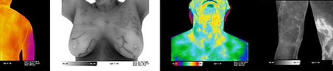
Medical Thermography uses an infrared camera to record temperature variations on the skin – providing information directly influenced by vascular activity below the surface via the sympathetic nervous system. Visible heat patterns of activity vary in distribution over regions of the body. A physician can identify anomalies in the physiology from these heat patterns, including the presence of inflammation or infection.
Asymmetry Analysis
While a healthy human body presents a symmetrical heat pattern, asymmetric changes visible in the heat patterns may be recognised by the physician as abnormal physiology or function. For example; a warm pattern visible on one breast but not the other could indicate more blood flow or physiological activity in that breast.
Angiogenesis
Angiogenesis is the natural formation of new blood vessels and plays a critical role in the growth of cancer because tumours cannot grow beyond a certain size without a blood supply. The resulting new blood vessels “feed” growing tumours with oxygen and nutrients, allowing the cancer cells to invade nearby tissue, or move throughout the body.
The formation of new blood vessels is visible on a thermogram because the warmth of the blood generates heat, revealing a change in the heat pattern. Also, in an asymmetric analysis the different heat patterns on opposite sides of the body would be clearly visible.
Comparison of changes over time
One of the advantages of Thermography is that any changes in the physiology are very apparent over time. An initial test can be compared to a later test taken months or even years later, and any visible changes in the heat pattern will contribute to the decision-making process regarding therapy, additional testing and eventual diagnosis of disease or illness.
Preventive Health – staying one step ahead
With regard to breast cancer, most women discover they have a problem with their breast health AFTER they develop symptoms. At this point they are one step behind and clearly at a disadvantage.
A thermogram can reveal a physiological change which may LATER result in disease or illness, giving the advantage back to the patient. The earliest possible indication of abnormalities allows for the earliest possible intervention – close monitoring with regular thermograms allows the patient to consider any factors that could be affecting their health.
Dense Breasts
Dense breasts have more non-fatty gland tissue compared to less dense breasts that have more fatty tissue. Dense breasts do not pose a health threat, are normally attributed to younger women and generally become less dense as the women get older. Currently the only way to identify dense breasts is with a mammogram – but unfortunately tumours and dense breast tissue are indistinguishable.
A thermogram is not restricted by dense breasts and therefore has advantages for younger women and women with dense breasts.